A series of photographs were taken using an optical microscope with low magnification. In these tests sample P1 was divided into two parts, respectively designated as P1a and P1b.
| Id | Date | Location | Area | Description | Amount | Depth |
|---|---|---|---|---|---|---|
| P1 | Jan 9 | on the ring | tens of cm2 | surface spots | about 100 g | 2 cm |
| P2 | Feb 17 | 4 m away from ring | tens of cm2 | small shovelful of loose soil | about 200 g | surface |
| Q1 | Jan 9 | on the ring | few cm2 | surface soil | few g | surface |
| Q2 | Jan 9 | on the ring | few cm2 | depth soil | few g | 2 cm |
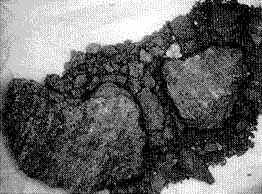
Sample P1a showed heavily compacted dirt with a crust 6 to 7 mm thick, predominantly composed of very dry limestone with only a few traces of dessicated vegetation in the form of moss. Curved striations are clearly seen on the surface, indicating that this dirt has been exposed to a rubbing effect that has resulted in the abrasion of some silicium grains (Figure 16). Further examination disclosed a spot where a small silex had been not only imprinted but ground to the level of the surrounding dirt (Figure 17). The soil has been fractured on either side of this silex, possibly under a combination of mechanical and thermal action. To the right of this area in Figure 17 it appears that the soil is darker and contains small vegetal shoots that have germinated after the gathering of the sample. The abrasion effect is less visible in that area.
Sample P1b comes from the same part of the ring as P1a. It exhibits similar compression effects as well as striations. It also shows a darker area that could correspond to foreign material or even to a transformation of the surface material (Figure 18). This is clearly observable in Figure 19 and also in Figure 20, where some plants are germinating and pushing back the black material.