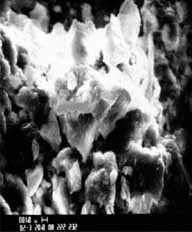
magnification 2,000.
size and shape, mostly ovoid or spherical. The particles range anywhere from 1.0 micrometer to 6.0 micrometers in size. In some fields there appear to be a few long tube-like structures protruding from the aggregates.
Again, these findings are consistent with the different depths of the material and the presence of biological material on the surface.
The samples identified as Q1 and Q2 were examined for elemental composition by energy dispersive x-ray analysis on the scanning electron microscope. More effort was directed toward the Q1 sample because of its greater interest to the scientists, due to its diversity. (Again, the scientists did not know the origin and nature of the samples and were only guided by their own deductions.)
The samples were analyzed using 20 keV electrons over several fields at both low and high magnification. Two other samples were examined to provide background information on common constituents of "dirt," which was the gross appearance of these samples.
Both samples contain aluminum, silicon, calcium, and iron. Sample Q1 also contains potassium in low concentration. The presence of sodium may